Reproducible Peptide MS/MS for Proteomics Labs: Lock Parameters
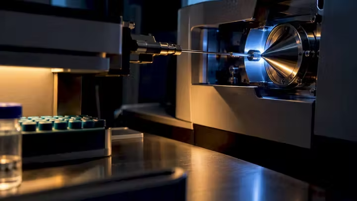
Peptide MS/MS analysis identifies and characterizes peptides by matching precursor mass, fragmentation spectra, and peptide-spectrum matches (PSMs) against a reference or de novo search space. The single most urgent action before trusting any result set: confirm your acquisition mode (DDA or DIA), verify the fragmentation method matches your search engine’s expected ion series, and check that your search space and decoy strategy are correctly configured. Skipping that check is how chimeric spectra and mismatched search parameters quietly inflate your false discovery rate.
TL;DR:
Confirm that your acquisition mode, fragmentation method, and search space are correctly configured to prevent inflating false discovery rates.
Use narrow isolation windows below 1.2 Da for cleaner spectra, but be aware they slow acquisition; select fragmentation methods based on expected ion series.
Document enzyme specificity, modifications, and search parameters thoroughly to ensure reproducibility and accurate peptide identification.
Choose your search strategy—database, open, or de novo—based on your sample type and expected modifications, and run multi-pass searches when necessary.
Archive all raw, converted, and result files, along with metadata, to enable full reproducibility and validation of your peptide analysis workflow.
Table of Contents
What Is MS/MS Analysis for Peptides and Why the Workflow Matters
Reading MS/MS Spectra: Fragment Ion Chemistry That Actually Matters
Database Search, Open Search, or De Novo: Choosing an Identification Strategy
DDA vs DIA: Acquisition Modes and Spectral Library Strategy
Search Engines, Rescoring, and DIA Software: Building a Reproducible Pipeline
Quantification Modes, Normalization, and Error Control
What to Archive So Others Can Reproduce Your Results
Research-Use Disclaimer and Where to Verify Your Standards
A Practical Decision Flow for Your Next Dataset
Author Perspective: The Fastest Way to Waste a Good Dataset
Sourcing Verified Peptides for Your Validation Workflow
Sources
FAQ
What Is MS/MS Analysis for Peptides and Why the Workflow Matters
MS/MS peptide identification depends on a chain of decisions that starts well before the mass spectrometer ever fires. Each stage in the pipeline, from digestion chemistry to search algorithm, shapes what you can and cannot say about your final PSM list. Get one link wrong and you either lose real identifications or accumulate false ones that look statistically clean.
The end-to-end workflow generally runs through six checkpoints:
Sample prep and digestion: trypsin remains the default enzyme for bottom-up proteomics because it cleaves predictably at lysine and arginine, producing peptides in the 700 to 3,000 Da range that fragment well. Lys-C, chymotrypsin, or dual-enzyme digestion strategies expand coverage for missed-cleavage-prone regions or when targeting specific PTM sites.
Peptide cleanup: solid-phase extraction or C18 desalting removes salts and detergents that suppress ionization. Skipping this step is a common source of poor spectral quality in student and early-career datasets.
LC separation: gradient length and column chemistry determine peak capacity. Longer gradients resolve more co-eluting peptides but cost instrument time.
MS acquisition settings: isolation window width, resolution, and cycle time all trade off identification depth against speed.
Vendor raw to mzML conversion: tools like ProteoWizard’s msConvert standardize proprietary vendor formats into open formats for downstream search engines.
Search and quant: this is where database search, open search, or de novo strategies assign identities to spectra.
Fragmentation method and isolation width matter more than most researchers give them credit for. A narrow isolation window (below 1.2 Da) reduces co-isolation of near-mass peptides but slows acquisition. Higher-energy collisional dissociation (HCD) versus electron-transfer dissociation (ETD) changes which fragment ion series you should even expect to see, which affects search engine scoring downstream.
Preserve your raw vendor files and mzML conversions together, along with mzIdentML or search-engine output tables. Losing the raw file means losing the ability to reprocess with an updated algorithm or corrected parameter set, a problem that surfaces more often than researchers expect when a reviewer asks for reanalysis.
Reading MS/MS Spectra: Fragment Ion Chemistry That Actually Matters
Fragment ion series are not interchangeable, and the activation method you choose determines which series you should be looking for. Collision-induced dissociation (CID) and HCD both cleave preferentially at the amide backbone, generating b-ions and y-ions. Electron-transfer dissociation and electron-capture dissociation instead cleave the N-Cα bond, producing c-ions and z-radical ions (c/z•), which behave differently under scoring algorithms tuned for b/y patterns.

Knowing which series to expect changes how you judge a candidate PSM. A HCD spectrum scored against a b/y model that shows dominant c/z• signal is a red flag for either mislabeled activation metadata or a data-processing error upstream.
Several recurring artifacts complicate spectral interpretation:
Neutral losses: water and ammonia losses from serine, threonine, or arginine residues create satellite peaks that can be misread as separate fragment ions.
Internal fragments: secondary cleavages within a peptide backbone generate ions that don’t fit the standard b/y or c/z ladder, and low-quality search engines sometimes force-fit them.
Isotopic envelope misassignment: picking the wrong isotope peak as the monoisotopic mass throws off the entire precursor mass calculation.
Co-fragmentation and chimeric spectra: two peptides co-isolated in the same window produce a mixed spectrum that neither peptide’s theoretical fragments fully explain, a frequent hidden failure mode in busy runs, as noted in reviews of bottom-up proteomics.
Before trusting a candidate identification, run through a short checklist: precursor mass error (should sit within your instrument’s calibrated tolerance, typically under 10 ppm on modern Orbitrap or time-of-flight systems), charge state consistency, isotope pattern shape, matched fragment ion coverage across the peptide backbone, and whether the relative intensity pattern matches what you’d expect from the activation method used.
Pro Tip: When a PSM looks borderline, manually recalculate at least the two or three most intense unmatched peaks using a fragment ion calculator. The Hunt Lab guide to de novo peptide sequencing walks through this by hand and it’s worth doing once on real data before you trust automated scoring blindly.
Database Search, Open Search, or De Novo: Choosing an Identification Strategy
The three dominant identification strategies solve different problems, and picking the wrong one for your sample type is one of the most common ways researchers lose real peptide identifications without ever knowing it.
Database search works best when your sample matches a well-annotated proteome and you can specify expected variable modifications up front. This is the default for most human, mouse, or yeast proteomics work. Its accuracy depends heavily on getting enzyme specificity, missed cleavage tolerance, and modification lists right before you run it. MSFragger’s fragment-indexing approach makes conventional database search fast even across large search spaces, and specifying frequently occurring modifications explicitly (rather than relying on open search to find them) measurably improves sensitivity for those modifications, according to the tool’s original methods paper.
Open search widens the precursor mass tolerance dramatically, often to several hundred daltons, to catch modifications you didn’t think to specify. This reveals unanticipated mass shifts, unusual PTMs, and chemical artifacts from sample handling. The tradeoff is that open search results need more careful post-processing and modification localization before you can treat a mass shift as biologically meaningful. MSFragger’s architecture was specifically built to make open searching computationally practical at scale.
De novo sequencing skips the reference database entirely and reconstructs peptide sequence directly from fragment ion spacing. This matters most for immunopeptidomics, noncanonical peptide discovery, and samples where no matching database exists. It comes with a real cost: de novo results lack a universally accepted FDR estimate, though frameworks like NovoBoard attempt decoy-spectrum strategies to approximate one. Treat de novo hits as hypotheses requiring orthogonal validation, not final answers.
Practical advice that applies across all three strategies:
Document enzyme specificity and missed cleavage settings explicitly in every search.
Record variable modification lists and precursor/fragment tolerances used.
Run multi-pass searches (a tight first pass, then a wider second pass on unmatched spectra) when your sample likely contains modifications you can’t fully predict in advance.
DDA vs DIA: Acquisition Modes and Spectral Library Strategy
Data-dependent acquisition (DDA) selects individual precursor ions in real time, usually the most intense ions in a survey scan, then fragments them one at a time. Data-independent acquisition (DIA) instead fragments broad, predefined mass windows regardless of what’s underneath them, then relies on computational deconvolution to assign fragment ions back to their originating precursors.
DDA gives you cleaner, more directly interpretable individual spectra, but it’s inherently biased toward abundant peptides and stochastic in which precursors get sampled run to run. DIA trades that clean-spectrum simplicity for comprehensive, reproducible coverage across replicates, at the cost of far more complex deconvolution.
That deconvolution step lives or dies on your spectral library. Three common strategies:
Empirical project-specific libraries: built from your own DDA runs on the same or similar samples, offering the tightest match to your actual instrument and chromatography.
Predicted in-silico libraries: generated computationally from sequence alone, useful when you lack matched DDA data but generally less precise for retention time prediction.
Gas-phase fractionation-refined libraries: built using narrow-window DIA runs specifically to deepen library coverage before the main quantitative experiment.
Benchmark studies comparing DIA software and library combinations show that your choice materially changes both identification counts and quantitative accuracy, not just at the margins. A comprehensive benchmarking study of DIA workflows found meaningful performance differences tied directly to library type and software settings, which means the “right” combination for one lab’s clinical samples may underperform for another lab’s cell culture work. Run a pilot comparison on representative samples before committing your full study to one library strategy. It’s cheaper to discover a mismatch on 10 pilot runs than on 200 production runs.
Search Engines, Rescoring, and DIA Software: Building a Reproducible Pipeline
A handful of tools now cover most of the peptide identification landscape, and each occupies a distinct role rather than competing head-to-head.
MSFragger, run inside the FragPipe environment, handles the core database and open search step. Its fragment-indexing architecture makes it fast enough to run open searches routinely, and FragPipe wraps it with PTM discovery modules and downstream quantification tools.
Percolator takes PSM candidates and rescoring features and applies semi-supervised SVM learning to separate correct from incorrect matches more precisely than a single score threshold ever could.
MSBooster sits between MSFragger and Percolator, generating predicted retention time, ion mobility, and MS2 spectral features for each candidate peptide, then feeding those predictions into Percolator as additional rescoring evidence. Integrating MSBooster into a FragPipe plus Percolator pipeline has been shown to increase identified PSMs and peptides at a fixed FDR, according to the tool’s methods paper, because it gives the rescoring model more independent evidence to work with than spectral matching alone provides.
DIA-NN handles DIA-specific deconvolution and quantification, working from either empirical or predicted spectral libraries.
Skyline provides a flexible interface for targeted quantification, chromatogram review, and manual verification across both DDA and DIA data types.
Three pipeline templates cover most research needs:
Identification and PTM discovery: FragPipe (running MSFragger) plus MSBooster plus Percolator, tuned for open search when modification discovery is the goal.
DIA quantification: DIA-NN paired with either a refined empirical library or an in-silico predicted library, benchmarked on pilot samples first.
Label-free quantification: FragPipe feeding into IonQuant for MS1-level quantitative comparison across samples.
Pro Tip: Enable DL-based rescoring selectively, not by default on every run. It adds real value on complex or low-abundance samples but can also add processing time you don’t need on straightforward, high-confidence database searches. Always capture exact software version numbers and parameter files alongside your results.
Quantification Modes, Normalization, and Error Control
Choosing a quantification mode depends on your study design, not just instrument capabilities. MS1-level label-free quantification works well for straightforward abundance comparisons without added cost. Labeled approaches like TMT or iTRAQ multiplex samples in a single run, improving throughput and reducing batch effects, with tradeoffs in complexity and dynamic range. Fragment-level DIA quantification uses MS2 ion intensities, often improving specificity.
Whichever mode you choose, the statistical foundation matters as much as the acquisition choice. Best practices include:
Normalize across runs using median or quantile methods before comparing abundances.
Design adequate biological and technical replicates before you acquire data, not after you see disappointing p-values.
Handle missing values explicitly, whether through imputation or by modeling missingness as informative, rather than silently dropping rows.
Apply batch correction when samples span multiple acquisition days or instrument recalibrations, a factor quantitative workflow reviews flag as an underappreciated source of false positives.
FDR itself needs to be reported at the correct level. Target-decoy strategies, where reversed or scrambled sequences estimate the rate of incorrect matches, remain the standard method for controlling false discovery in large-scale searches. But PSM-level FDR and peptide-level FDR are not interchangeable numbers.
Peptide-level procedures, particularly PSM-and-peptide competition approaches, have been shown to yield more peptide discoveries at a fixed 1% FDR than PSM-only filtering, giving you more statistical power without loosening your actual error tolerance.
Always state explicitly whether you filtered at the PSM, peptide, or protein level. For any PSM central to your paper’s conclusions, manually inspect the spectrum and, where feasible, confirm with an orthogonal method like a synthetic peptide standard or targeted MRM.
What to Archive So Others Can Reproduce Your Results
Reproducibility failures in peptide MS/MS analysis rarely come from bad instruments. They come from missing records. A defensible analysis archive should include the raw vendor files themselves, the converted mzML copies, the exact FASTA database version and its provenance, complete search parameter files, software version numbers for every tool in the pipeline, the decoy generation method used, and the final filtered result tables.
Metadata worth recording alongside the files:
Instrument model and firmware version
Fragmentation method (HCD, CID, ETD, or a combination) and collision energy settings.
Isolation window width for DIA runs
LC gradient length and column chemistry
Sample prep notes, including enzyme, digestion time, and cleanup method
A complete record, covering everything from raw files through decoy strategy and FDR levels, is what separates a result a peer can rebuild from one they simply have to trust. Repository platforms like ProteomeXchange and PRIDE exist specifically to host this kind of archive publicly, and increasingly, journals require it as a condition of publication.
Research-Use Disclaimer and Where to Verify Your Standards
Every peptide discussed in this article is described strictly as a research compound intended for laboratory use. Nothing here constitutes dosing, administration, or reconstitution guidance for human or animal use, and none of it should be interpreted that way.
If your quantitative workflow depends on labeled internal standards, understanding how isotopically labeled peptides function in a quant pipeline is worth a closer look before you order material. And because search-engine output is only as trustworthy as the input material behind it, verifying purity and identity on any peptide standard you source matters just as much as your search parameters. Batch-specific certificates of analysis alongside third-party testing results are an important practice to expect from any supplier feeding your validation pipeline.
A Practical Decision Flow for Your Next Dataset
Skip the instinct to rerun everything when results look off. Start with quick QC: check total identifications, median mass error, and missed cleavage rate against what you’d expect for your sample type. If those look reasonable, move to a parameter check, confirming enzyme, modifications, and tolerances actually match your experiment before touching the raw data again.
Only reconfigure search parameters if the QC and parameter check both point to a setup mismatch. Re-acquire data only when the spectra themselves are the problem, not the search settings. And lock your software versions and parameter files the moment you’re satisfied with a result. Documentation written after the fact is documentation that gets details wrong.
Author Perspective: The Fastest Way to Waste a Good Dataset
The mistake I see most often isn’t a bad instrument or a weak search engine. It’s researchers treating acquisition mode and search strategy as afterthoughts instead of the first decision in the pipeline. A DDA dataset searched with DIA-tuned assumptions, or a database search missing an obvious modification, produces confident-looking output that’s simply wrong.
The gap between what a search engine reports and what’s actually true in your sample rarely comes from the algorithm itself. FragPipe, MSFragger, Percolator, DIA-NN, Skyline, and MSBooster are all mature, well-validated tools. The gap comes from parameter mismatches nobody checked: the wrong fragmentation assumption, an unspecified common modification, a decoy strategy nobody documented. Fix the input decisions first. The software will do its job correctly once you do yours.
If there’s one habit worth adopting from this entire workflow, it’s locking your parameter files and software versions before you write a single conclusion. Reanalysis six months later, when a reviewer asks a hard question, should take an afternoon, not a week of reconstructing what you actually ran.
— Adrian K. Solis
Sourcing Verified Peptides for Your Validation Workflow
Research-grade peptides with batch-specific analytical verification, including HPLC and mass spectrometry data, help ensure the material feeding your MS/MS pipeline is documented from the start.
Confident peptide identification only means something if the reference material behind it is what the label says it is. Transparent laboratory partnerships and published certificates of analysis for every batch support researchers in taking purity with confidence. That matters most when you’re running standards for quantitative work or validating a novel PTM finding against a known reference. Browse the Research Peptides category for verified standards, check Peptide Stacks for combined-compound options, and pick up Bacteriostatic Water for lab handling needs. Place your order today and get batch documentation alongside your shipment.
FAQ
How Do I Interpret LC-MS/MS Results?
Interpretation starts with the PSM list, not the raw spectra: check precursor mass error, matched fragment coverage, and charge state consistency for each candidate before trusting the identification. Then confirm which FDR level (PSM, peptide, or protein) was applied, since peptide-level filtering typically yields more discoveries at the same 1% FDR than PSM-only filtering.
How Much Does a Proteomics Analysis Typically Cost?
There’s no single standard price, since cost depends heavily on sample prep complexity, instrument time, fractionation needs, and whether the work runs in-house or through a core facility, a variability proteomics cost reviews confirm directly. Request a scoped quote from your core facility or vendor rather than relying on a generic figure.
What Is LC-MS/MS Analysis of Peptides and How Does It Work?
LC-MS/MS separates peptides by liquid chromatography, then fragments each precursor ion in the mass spectrometer to generate a spectrum matched against a database, an open search space, or reconstructed de novo. The output is a set of PSMs, which then get filtered by FDR before being interpreted as confident peptide or protein identifications.
What Is LC-MS/MS Analysis Used For?
It identifies and quantifies peptides and proteins in complex biological samples, supporting everything from PTM mapping to biomarker discovery to immunopeptidomics. Researchers also use it to validate synthetic peptide identity and purity, which is why batch-specific MS verification matters when sourcing reference material for a study.
Why Does Fragmentation Method Change How I Read a Spectrum?
CID and HCD activation produce primarily b-ion and y-ion series, while ETD and ECD generate c-ion and z-radical ion series, so a search engine tuned for the wrong series will misscore correct matches. Always confirm your acquisition metadata records the actual activation method used before troubleshooting a poor-scoring PSM.
Recommended