Four Stages of Peptide Mass Spectrometry and COA Checks for Labs
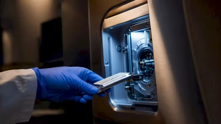
Mass spectrometry gives peptide researchers three things no other technique delivers together: precise molecular mass, sequence-level confirmation through fragmentation, and localization of post-translational modifications. The standard approach is bottom-up LC-MS/MS, where peptides are separated by liquid chromatography and analyzed by tandem mass spectrometry, benchmarked against resources like NIST’s peptide spectral libraries and de novo tools from the Hunt Lab. Some peptide distributors rely on this same analytical backbone to verify research-grade peptides.
TL;DR:
Peptide mass spectrometry’s key advantages include simultaneous precise mass measurement, sequence confirmation, and post-translational modification localization.
Proper sample cleanup and verified starting materials are critical, as residual impurities and unconfirmed reagents can severely compromise spectra quality.
Hybrid instruments combining quadrupole and Orbitrap analyzers are common for balancing speed, resolution, and confident identification, especially in PTM mapping.
Fragmentation methods like ETD or ECD are essential for accurate PTM localization and sequencing of longer, highly charged peptides.
Using verified research-grade peptides with batch-specific certificates of analysis significantly reduces false positives and improves experimental reliability.
Table of Contents
What Are the Four Stages of Peptide Mass Spectrometry?
How Do You Prepare Peptide Samples for Mass Spectrometry?
Which Ionization Method and Mass Analyzer Should You Use?
What Do CID, HCD, and ETD Fragmentation Reveal?
Should You Choose DDA or DIA for Peptide Quantification?
How Does Peptide Identification Software Work?
How Do You Troubleshoot Low-Signal or Noisy MS Runs?
Why Does Peptide Verification Matter for Mass Spectrometry Results?
The Editorial Take: Verification Beats Instrumentation Every Time
Get Verified Research-Grade Peptides From Rapidcorebio
Sources
FAQ
What Are the Four Stages of Peptide Mass Spectrometry?
Every peptide mass spectrometry workflow, regardless of instrument brand or lab, breaks down into four operations performed by the MS system itself: creating ions from your peptide sample, separating those ions by mass-to-charge ratio (m/z), detecting them, and selectively fragmenting precursor ions to generate sequence-informative MS/MS spectra. This four-part structure comes straight from the foundational proteomics literature at the Broad Institute, and it holds true whether you are running a quadrupole time-of-flight instrument or an Orbitrap.
Where researchers get tripped up is choosing the right strategy for the biological question. Three approaches dominate peptide-level proteomics, and they are not interchangeable:
Bottom-up proteomics digests proteins into peptides (usually with trypsin) before analysis. This is the workhorse approach for large-scale identification and PTM mapping, and it is what most people mean when they say “peptide mass spectrometry.”
Top-down proteomics analyzes intact proteins without digestion, preserving the full modification pattern but sacrificing throughput and sensitivity for larger proteins.
Middle-down proteomics splits the difference, using limited proteolysis to generate larger peptide fragments that retain more modification context than a fully digested bottom-up sample.
Liquid chromatography runs before the mass spectrometer for a practical reason: a tryptic digest of even a modest protein mixture produces thousands of peptides, and no mass analyzer can resolve them all if they hit the ion source simultaneously. LC separates peptides by hydrophobicity over a gradient, feeding the mass spectrometer a manageable stream instead of a chemical traffic jam. Trypsin remains the default digestion enzyme because it cuts predictably after lysine and arginine residues, generating peptides in a range generally suitable for most instruments. Chymotrypsin, Lys-C, and Glu-C serve as alternatives when you need different cleavage specificity or better coverage of a particular sequence region.
How Do You Prepare Peptide Samples for Mass Spectrometry?
Sample preparation is where most peptide MS experiments succeed or fail, long before any ion reaches the detector. Practitioner guidance consistently identifies inadequate cleanup as the single most common source of failed or noisy peptide spectra, and the mechanism is straightforward: residual salts, detergents, and polymers compete with your peptide for charge during ionization, suppressing the signal you actually want to measure.
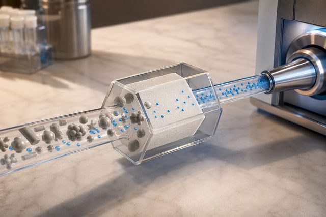
Enzymatic digestion with trypsin typically runs overnight at a controlled temperature, following denaturation and reduction/alkylation steps that unfold the protein and break disulfide bonds so the enzyme can access cleavage sites. Skipping or shortening this step leaves missed cleavages that complicate downstream identification. Once digestion is complete, cleanup becomes the priority. Solid-phase extraction (SPE) and StageTips, small pipette-tip-format C18 cartridges, are the two most common desalting approaches, both working on reverse-phase principles to bind peptides while salts and buffer components wash through. Concentration by vacuum centrifugation or lyophilization follows, ideally without allowing samples to fully dry out and become difficult to resuspend.
Ion suppression deserves specific attention because it is often invisible until you are staring at a chromatogram with almost no peaks. Common culprits include:
Residual salts (phosphate buffers, Tris) from lysis or digestion buffers, fixed by additional desalting passes.
Detergents like SDS or Triton X-100 used in cell lysis, which persist through inadequate cleanup and devastate ionization efficiency.
Polymer contamination from plastic labware or gloves, identifiable by characteristic repeating mass peaks in the spectrum.
Keratin contamination from skin flakes or hair, a near-universal problem that shows up as an unwanted background signal in low-abundance samples.
Plasticizers and lubricants leaching from pipette tips or tubes, worth ruling out when background noise is unexpectedly high.
Before loading any sample onto the instrument, run through a short quality-control pass: confirm digestion completeness with a quick check for missed cleavages, verify peptide concentration against a standard curve, inspect for visible particulate, and confirm your solvent gradient and mobile phases are fresh and free of microbial growth (a surprisingly common issue with aqueous buffers left too long at room temperature).
Pro Tip: Every time you transfer a peptide sample between tubes, you lose material to the plastic surface through nonspecific adsorption. Minimize the number of transfers between digestion and injection, and use low-binding tubes for dilute samples. This single habit change often recovers more signal than an extra round of desalting.
Which Ionization Method and Mass Analyzer Should You Use?
Electrospray ionization (ESI) and matrix-assisted laser desorption/ionization (MALDI) are the two soft ionization techniques that dominate peptide mass spectrometry, and they solve different problems. ESI is the standard partner for LC-MS because it ionizes peptides directly from liquid eluent, making it the natural choice whenever chromatographic separation precedes mass analysis. MALDI ionizes samples embedded in a solid matrix, which suits high-throughput peptide mapping and situations where speed matters more than the deep separation LC provides.
Choosing a mass analyzer is really a question about what you are trying to learn. The trade-offs are well established:
Quadrupole analyzers filter ions by m/z with speed and precision, making them the go-to choice for targeted quantification of known peptides rather than open-ended discovery work.
Ion traps excel at MSn experiments, where you fragment an ion, then fragment the resulting fragment again, useful for deep structural characterization of a single peptide of interest.
Time-of-flight (TOF) analyzers measure how long ions take to travel a fixed distance, offering fast scan speeds well suited to high-throughput screening.
Orbitrap analyzers trap ions in an electrostatic field and measure their oscillation frequency, delivering the resolution and mass accuracy that modern PTM mapping and complex mixture analysis demand.
Fourier-transform ion cyclotron resonance (FTICR) analyzers push resolution even further using a magnetic field, at the cost of longer analysis times and higher instrument complexity.
Most contemporary proteomics labs do not pick just one. Hybrid instruments pairing a quadrupole front end with an Orbitrap back end have become the default configuration for a reason: the quadrupole handles fast precursor selection and low-resolution filtering, while the Orbitrap delivers the high-resolution mass measurement needed for confident peptide identification. This combination lets you run quantitative and qualitative analysis in the same acquisition without compromising on either speed or accuracy.
What Do CID, HCD, and ETD Fragmentation Reveal?
Fragmentation is where a peptide’s mass measurement turns into an actual sequence. Collision-induced dissociation (CID) and higher-energy collisional dissociation (HCD) both work by colliding peptide ions with an inert gas, breaking the peptide backbone predominantly at the amide bond to generate b-ions and y-ions. HCD has become the preferred method on Orbitrap instruments specifically because it produces fragments compatible with high-resolution detection in the same analyzer, whereas classic CID on ion traps sometimes requires a separate detection step. Both methods share a known weakness: labile modifications like phosphorylation frequently fall off as neutral losses before the backbone fragments, which can obscure exactly where a modification sits.
Electron-transfer dissociation (ETD) and electron-capture dissociation (ECD) solve that problem by breaking the peptide backbone through a different chemical mechanism, generating c-ions and z-ions while leaving labile PTMs intact on the fragment where they belong. This makes ETD and ECD the preferred choice when the research question centers on PTM localization or when sequencing longer, more highly charged peptides where CID/HCD fragmentation becomes uneven.
A few specialized techniques round out the fragmentation toolbox:
Surface-induced dissociation (SID) collides ions with a solid surface instead of gas molecules, producing more uniform fragmentation energy useful for structural studies of larger complexes.
Infrared multiphoton dissociation (IRMPD) uses laser energy for a gentler, tunable fragmentation process, often paired with FTICR instruments.
Combining complementary fragmentation types (running both HCD and ETD on the same precursor) reliably improves sequence coverage compared to relying on a single method.
Pro Tip: If your sequence coverage looks incomplete around a known modification site, don’t just increase collision energy. Switch fragmentation mode instead. An ETD run on the same precursor often recovers exactly the c/z ions that HCD neutral-loss chemistry destroyed.
Should You Choose DDA or DIA for Peptide Quantification?
Data-dependent acquisition (DDA) has been the default acquisition strategy in peptide mass spectrometry for years, and understanding its limitation is critical to interpreting your own results. DDA selects the most abundant precursor ions in real time for fragmentation, which means lower-abundance peptides are stochastically sampled. Run the same sample twice and you will not fragment an identical set of precursors both times, a real constraint for discovery experiments trying to catalog every peptide present in a complex mixture.
Data-independent acquisition (DIA) takes a fundamentally different approach, fragmenting all precursor ions within defined m/z windows regardless of abundance. The resulting MS/MS spectra are complex composites of multiple co-fragmented peptides, which requires either a pre-built spectral library or library-free computational deconvolution to assign fragments back to their correct precursor. DIA trades acquisition simplicity for a harder downstream computational problem, but it delivers far more reproducible quantification across sample sets.
Quantification strategy is a separate decision layered on top of acquisition mode:
Label-free quantification compares peptide peak areas or spectral counts directly across separate LC-MS/MS runs, requiring no chemical labeling but demanding tight run-to-run reproducibility.
Isobaric tagging (TMT or iTRAQ) chemically labels peptides from different samples with mass tags that are identical in precursor mass but generate distinct reporter ions upon fragmentation, allowing multiple samples to be pooled and run together.
Choose label-free when sample number is small and instrument time is not the limiting factor; choose isobaric tags when you need to multiplex many samples and minimize run-to-run variation.
Neither acquisition strategy nor quantification method is universally correct. A discovery experiment cataloging an unknown peptide population usually starts with DDA. A study tracking the same 200 known peptides across 40 samples is a better fit for DIA or TMT multiplexing.
How Does Peptide Identification Software Work?
Turning raw spectra into a confident peptide identification runs through one of three computational approaches, each suited to a different scenario. Database search engines compare experimental MS/MS spectra against theoretical fragmentation patterns generated from a protein sequence database, the standard choice when you are analyzing a well-characterized organism’s proteome. Spectral-library search matches experimental spectra directly against previously observed, validated spectra, an approach the NIST peptide mass spectral libraries support directly. De novo sequencing algorithms reconstruct a peptide sequence from fragment mass differences alone, without any database assumption, which makes them essential for detecting novel peptides, unexpected modifications, or sequences from organisms lacking a complete reference proteome.
False discovery rate (FDR) control is what keeps any of these methods honest. Standard practice searches spectra against a decoy database, typically a reversed or randomized version of the real one, and calculates FDR from the ratio of decoy matches to real matches at a given confidence threshold. A 1% FDR cutoff is conventional in most peptide identification pipelines, meaning roughly one in a hundred accepted identifications is expected to be a false positive.
Mass accuracy measured in parts per million is one of the primary drivers of false-positive reduction in peptide database searches, because tighter mass tolerance windows exclude far more incorrect candidate matches before scoring even begins.
Low-ppm accuracy matters because several amino acid combinations produce near-identical masses at coarse resolution, and only tight mass windows reliably distinguish them. This is precisely why Orbitrap and FTICR instruments, with their superior mass accuracy, have become standard for confident large-scale peptide identification rather than a luxury upgrade.
De novo sequencing remains the fallback when database and spectral-library searches come up empty, and it is not a niche skill. The Hunt Lab’s tutorials and fragment calculators are widely used training resources for manual MS/MS interpretation, and deep-learning models published in 2024 have meaningfully improved de novo sequencing sensitivity and precision on difficult datasets, narrowing the gap between automated de novo calls and expert manual annotation.
Key tool categories to know:
Database search engines for standard proteome-matched identification.
Spectral-library search tools for validated, previously characterized peptides.
De novo sequencing algorithms, increasingly deep-learning assisted, for novel sequence discovery.
Decoy-based FDR calculators built into most modern search pipelines as a standard output.
How Do You Troubleshoot Low-Signal or Noisy MS Runs?
When a run produces weak or unexpected spectra, work through the problem systematically rather than re-injecting the same sample and hoping for a different result.
Check solvent and mobile phase quality first. Aged buffers, microbial growth, or an improperly mixed gradient are common and easy to overlook.
Confirm the sample was desalted adequately. If ion suppression is suspected, run a quick desalting pass on a split of the original sample before assuming the peptide itself is degraded.
Inspect the nano-ESI emitter for clogs or damage. A partially blocked emitter produces a slow, unstable spray that looks like a chemistry problem but is a hardware one.
Verify instrument calibration against a known standard. Mass accuracy drifts over time, and a stale calibration silently degrades identification confidence.
Re-examine low-confidence peptide-spectrum matches (PSMs) manually rather than discarding them outright. A PSM that fails automated scoring by a narrow margin sometimes reflects a real peptide with an unanticipated modification the search parameters did not include.
When standard search pipelines still fail to explain a spectrum, experienced labs turn to custom scripts or manual annotation tools that account for non-standard adducts or lab-specific modifications the default software was never configured to detect. This kind of hands-on correction is routine at labs doing frontline PTM discovery work, not a sign that something went wrong with the experiment.
Pro Tip: Keep a running log of instrument calibration dates alongside your sample run log. When results look inconsistent across a project, the first cross-check should be whether a calibration drift coincides with the affected runs, not whether the biology changed.
Why Does Peptide Verification Matter for Mass Spectrometry Results?
A confident peptide identification depends on starting material you can trust, and this is where analytical verification earns its place in the workflow rather than serving as a formality. A certificate of analysis (COA) for a research peptide typically reports the compound’s identity confirmed by mass spectrometry, purity determined by HPLC, and a lot number tying the results to the specific material in hand. Without that documentation, an unexpected mass shift or extra peak in your own MS run is ambiguous: is it a genuine modification, a synthesis byproduct, or degradation from poor storage?
Verified starting material removes one entire category of confounding variable from your spectra. When you already know the peptide’s expected mass and purity from independent HPLC and MS analysis, an anomalous peak in your own experimental data points you toward sample handling or instrument conditions rather than sending you chasing a phantom impurity that was in the vial from the start. Worth requesting from any supplier before you commit instrument time to a batch:
The specific lot number’s COA, not a generic or representative one for the product line.
Both HPLC purity data and MS-confirmed identity, not one or the other.
Storage and stability guidance appropriate to the peptide’s chemistry, since improper storage before analysis can introduce degradation products that mimic real PTMs on a spectrum.
Rapidcorebio provides batch-specific COAs backed by third-party HPLC and mass spectrometry testing for its research-grade peptide catalog, along with a research handbook and guidance on sample handling techniques built for exactly this kind of pre-analysis diligence. Researchers comparing peptide and protein behavior in MS workflows, or working with isotopically labeled standards for quantification and mass accuracy checks, will find the same verification logic applies directly to calibration and internal standard selection.
The Editorial Take: Verification Beats Instrumentation Every Time
Most guides to peptide mass spectrometry obsess over instrument choice, as if the difference between an Orbitrap and a quadrupole is where experiments go wrong. It rarely is. The failure point researchers underestimate is almost always upstream: a poorly desalted sample, an unverified starting reagent, or a calibration nobody rechecked in weeks. You can own the best hybrid quadrupole-Orbitrap on the market and still generate garbage spectra from a sample carrying residual detergent.
The conventional advice on this topic treats sample prep and reagent sourcing as boring prerequisites to get through quickly before the “real” analytical work begins. That framing has it backwards. Sample prep and starting material quality determine what your instrument is even capable of showing you. A researcher who skips COA verification to save a few days is not saving time. They are importing an unknown variable into every spectrum they will spend the next month interpreting.
If you take one thing from this guide, prioritize verification before acquisition mode. Confirm what is in the vial, confirm your desalting worked, then argue about DDA versus DIA.
— Adrian K. Solis
Get Verified Research-Grade Peptides From Rapidcorebio
Rapidcorebio removes the biggest unknown in peptide mass spectrometry work: whether your starting material is actually what the label says. Every batch ships with a research-grade certificate of analysis backed by third-party HPLC and MS testing, so an unexpected peak in your spectra points you toward your own workflow instead of an unverified reagent.
Beyond the COA page, Rapidcorebio’s research handbook and peptide sampling technique guide walk through the handling steps that most directly affect MS signal quality, from digestion through storage. Labs building out calibration standards or PTM mapping panels can review Kisspeptin-10 product and other catalog peptides alongside their batch-specific COAs before ordering. If you have MS-specific questions about a particular lot, reach out to Rapidcorebio’s support team and verify the certificate of analysis for your exact batch before it goes anywhere near your instrument.
Sources
The Hunt Lab Guide to De Novo Peptide Sequence Analysis by Tandem Mass Spectrometry
Fundamentals of Biological Mass Spectrometry and Proteomics (Broad Institute PDF)
Nature article on deep learning for de novo peptide sequencing (2024)
This article covers research methodology only. Rapidcorebio’s peptides are sold strictly for laboratory research use and are not intended for human or animal consumption.
FAQ
What Does Mass Spectrometry Tell You About a Peptide?
Mass spectrometry provides the peptide’s precise molecular mass, sequence information through MS/MS fragmentation, and the location of post-translational modifications, typically through bottom-up LC-MS/MS analysis.
Is ESI or MALDI Better for Peptide Analysis?
ESI is the standard choice when peptides are separated by liquid chromatography beforehand, while MALDI suits high-throughput peptide mapping where speed matters more than upstream separation.
Why Does Sample Cleanup Matter So Much for Peptide MS?
Residual salts, detergents, and polymer contaminants suppress ionization and can damage nano-ESI emitters, making desalting one of the most common points of failure in peptide mass spectrometry.
What Is the Difference Between DDA and DIA?
DDA fragments the most abundant precursor ions in real time, which introduces run-to-run variability, while DIA fragments all precursors in defined m/z windows for more reproducible quantification at the cost of more complex data analysis.
How Does a COA Help With Mass Spectrometry Results?
A certificate of analysis confirms a peptide’s identity and purity through independent HPLC and MS testing, which helps researchers distinguish real experimental findings from artifacts introduced by unverified starting material; Rapidcorebio provides batch-specific COAs for this purpose.
Recommended